Jan
20
to Jan 24
WBIR 2024 Workshop
Sunday 6th October 2024, workshop fully in presence in Marrakesh, Morocco (no hybrid, online part)
In association with the MFS2023 Meeting, the MultEMplex CIG will have a two-day meeting in Braga on September 26 and 27th to discuss in detail the needs, constraints and the proposed design for the new EM-grid and workflow. Portions of the meeting are open to anyone in the electron microscopy community, and all MFS2023 participants are invited to join the open sessions and contribute to the discussions.
The MultEMplex CIG will cover venue and catering costs. However, advance registration is required, and a participation signature will be needed to verify your participation.
https://www.mfs2023.com/cig
This training school is sponsored by COMULIS as a COST Action (www.comulis.eu): COST (European Cooperation in Science and Technology) is a funding agency for research and innovation networks. Financial support to a limited number of participants available. Registration fee: 250 EUR Registration link: here
https://www.ana.unibe.ch/continuing_education/comulis_training_school/index_eng.html.
We are pleased to announce an exciting new lecture series brought to you by COMULIS and Euro-BioImaging as part of the Virtual Pub. Every third Friday of the month, starting in July 2021, the Virtual Pub will host speakers involved in the COMULIS COST Action for the “Correlative Imaging Series”. Talks will focus on multi-modality imaging combining a range of different imaging techniques and the biological and biomedical applications they can address.
Click here to join the talk.
Abstract:
Retina deposits form with age and their presence is the highest known risk factor for the development of age-related macular degeneration (AMD); the leading cause of blindness in older adults. Deposits, such as drusen and basal laminar deposits, form below the retinal pigmented epithelium (RPE) whereas deposits, such as subretinal drusenoid deposits (SDD), form above the RPE. The molecular composition of such deposits, which are only partly known, varies depending on their regional and laminar locations. Thus, accurate localization is essential for interpreting imaging mass spectrometry (IMS) results. Furthermore, given the exquisite architecture of the retina, a multimodal imaging approach is required to establish the localization of IMS signals. The general goal of this study is to use multimodal imaging methods including ex vivo OCT, autofluorescence, optical microscopy, and state-of-the-art IMS methods to elucidate the molecular composition of different retinal deposits in aged human tissue. Such information can molecularly inform clinical imaging modalities.
About Kevin Schey:
Kevin L. Schey, Ph.D, is Professor of Biochemistry, Chemistry, and Ophthalmology & Visual Sciences at Vanderbilt University. He earned his Ph.D. from Purdue University in 1989 and has 30+ years of professional experience in mass spectrometry and analytical chemistry, including all aspects of proteomics analysis and mass spectrometry imaging. His application area of interest is the eye, particularly lens and retina biochemistry. He directs the Core Facility operations of the MSRC.
Friday, May 20th, at 13:00 CET
Join via internet:
https://us02web.zoom.us/j/760003029
or via phone:
Meeting ID: 760 003 029
Find your local number: https://zoom.us/u/acj97wMY13
We are pleased to announce an exciting new lecture series brought to you by COMULIS and Euro-BioImaging as part of the Virtual Pub. Every third Friday of the month, starting in July 2021, the Virtual Pub will host speakers involved in the COMULIS COST Action for the “Correlative Imaging Series”. Talks will focus on multi-modality imaging combining a range of different imaging techniques and the biological and biomedical applications they can address.
Click here to join the talk.
Abstract:
Correlative light and electron microscopy (CLEM) entails a group of multimodal imaging techniques that are combined to pinpoint to the location of fluorescently labelled molecules in their ultrastructural context. Correlative super resolution and electron microscopy is one CLEM modality in which super resolution microscopy is used instead of conventional fluorescence microscopy techniques.
Single-molecule localization microscopy (SMLM) is one of the super resolution microscopy families, offering excellent resolution (5–25 nm), multi-colour imaging and quantification capability with single-particle precision. Thus, the improved resolution of SMLM leads to a nanoscale localization precision of the specific fluorescent labels in the ultrastructural reference space provided by EM.
Super-resCLEM methods have been mainly applied to biological samples; here we introduce it for synthetic materials. The decoration of nanoparticles with functional moieties is a key strategy to achieve cell targeting in nanomedicine. The interplay between size and ligand number is crucial for the formulation performance and needs to be properly characterized to understand nanoparticle structure−activity relations. However, there is a lack of methods able to measure both size and ligand number at the same time and at the single particle level. In this lecture, Silvia Pujals will address this issue by explaining how she and her team have introduced a super-resCLEM method, specifically by combining one type of SMLM (DNA-PAINT) with TEM.
Moreover, they are now applying super-resCLEM methods to learn about the trafficking of different nanomaterials inside cancer cells.
Andrian, T., Delcanale, P., Pujals, S.*, Albertazzi, L. Correlating Super-Resolution Microscopy and Transmission Electron Microscopy Reveals Multiparametric Heterogeneity in Nanoparticles. Nano Lett. (2021) 21(12):5360-5368.
About Silvia Pujals
Dr. Pujals is a Ramón y Cajal researcher at the Institute for Advanced Chemistry of Catalonia (IQAC-CSIC). She is also Adjunct Professor at the Department of Electronics and Biomedical Engineering, Faculty of Physics, Universitat de Barcelona.
She obtained her PhD in Organic Chemistry from Universitat de Barcelona (UB) in the field of cell-penetrating peptides. Then she moved to Kyoto University for her postdoc focused on biophysics and electron microscopy. Coming back to Barcelona she became senior researcher at the Nanoscopy for Nanomedicine group at IBEC, working on super resolution microscopy for nanomaterials. With expertise on drug delivery, peptide synthesis and optical and electron microscopy her research aims to combine a rational design of nanomaterials with advanced optical techniques for targeted drug delivery. "
Friday, March 18th, at 13:00 CET
Abstract:
Multimodal image datasets can comprise several TB of hundreds of images of different spatial resolution and different spatial extent. In addition, there can be thousands of objects that have been segmented in these images. To address the challenge of efficient scientific exploration of such datasets we developed MoBIE, a free and open-source platform for multimodal big image data exploration and sharing. MoBIE is an easy to install Fiji plugin, leveraging the lazy evaluation framework provided by ImgLib2 and BigDataViewer for smooth browsing of TB sized data sets on a standard laptop computer. Both file system and object store backends for image data hosting are supported as well as GitHub based storage of tabular data. MoBIE provides rich functionality for segmentation exploration as well as an expressive JSON schema for the specification of various views on a complex data set. We will also discuss the interplay of MoBIE with the upcoming OME.Zarr file format.
About Christian Tischer
Christian Tischer studied physics in Heidelberg, followed by PhD at EMBL with Philippe Bastiaens working on microscope development and signalling in mammalian cells. Subsequently, he did a postdoc with Marileen Dogertom at AMOLF in the Netherlands working on microtubule dynamics. From 2009 until 2018, he worked at EMBL's Advanced Light Microscopy Facility (ALMF) supporting scientists with both microscopy and image analysis. Since end of 2018, Christian’s focus shifted completely towards image analysis support and he is now running EMBL’s Centre for Bioimage Analysis (CBA).
Click here to join the talk.
We are pleased to announce an exciting new lecture series brought to you by COMULIS and Euro-BioImaging as part of the Virtual Pub. Every third Friday of the month, starting in July 2021, the Virtual Pub will host speakers involved in the COMULIS COST Action for the “Correlative Imaging Series”. Talks will focus on multi-modality imaging combining a range of different imaging techniques and the biological and biomedical applications they can address.
Click here to join the talk.
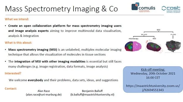
Mass spectrometry imaging (MSI) is an unlabeled, multiplex molecular imaging technique that allows the visualization of molecules in tissue sections. Integration with data from other imaging modalities is still challenging. In this meeting we want to discuss about this and start an open collaboration initiative between mass spectrometry imaging users and image analysis experts aiming to improve multimodal data sharing, visualisation, analysis & integration."
The last of four WG3 meeting to analyze the results obtained using the novel pipeline established within the COMULIS framework and finalize the publication
Further information will follow shortly
We are pleased to announce an exciting new lecture series brought to you by COMULIS and Euro-BioImaging as part of the Virtual Pub. Every third Friday of the month, starting in July 2021, the Virtual Pub will host speakers involved in the COMULIS COST Action for the “Correlative Imaging Series”. Talks will focus on multi-modality imaging combining a range of different imaging techniques and the biological and biomedical applications they can address.
Click here to join the talk.
Further information will follow shortly
Further information will follow shortly
During the last Grant Period, we recognized the importance of separate WG1 & 3 meetings to push forward novel multimodal pipelines in so-called showcase pipelines. This requires bringing together experts in diverse fields to work jointly on a biomedical research questions that will be tackled by correlative multimodal imaging including data integration and analysis. We plan to organize specific meetings for this, close to a laboratory which is already proactively imaging such a sample. AIm is to discuss our current showcase pipelines, analysis of previously implemented modalities, adjacent lab visits and sample exchange, and coordination of further steps. Developing a showcase project for CLEM & go beyond the established approaches, thus pushing the limits of CLEM. The meeting aims at setting up a pipeline that involves superresolution, which requires a broad expertise from diverse imaging scientists. Topics to coordinate and plan are (including the involvement of computational approaches and WG4): LGA-PEG Nanoparticles with STORM dyes encapsulated, incubated in MCF7 cells (breast cancer cell line, as Biliana is working with anticancer drugs for breast cancer). Live cell imaging (Confocal or SIM?). Fixation (controversial step!). Single molecule localization microscopy (STORM). Cryo fixation/resin embedding. Cryo-TEM/TEM. Image alignment
We are pleased to announce an exciting new lecture series brought to you by COMULIS and Euro-BioImaging as part of the Virtual Pub. Every third Friday of the month, starting in July 2021, the Virtual Pub will host speakers involved in the COMULIS COST Action for the “Correlative Imaging Series”. Talks will focus on multi-modality imaging combining a range of different imaging techniques and the biological and biomedical applications they can address.
Click here to join the talk.
Correlated Multimodal Imaging – enabling discoveries from atoms to anatomy
It is a great pleasure to welcome you to the Virtual Conference of COMULIS network 2021 in Gothenburg, Sweden. COMULIS 2021 is designed to bring experts, researchers, facility staff and exhibitors together from different imaging modalities to stimulate knowledge exchange and the formation of new collaborations. No matter where you are in your career, participating at the Conference will allow you to stay abreast of new correlated multimodal imaging technologies. Take the opportunity to challenge your brain, create a new network and maintain existing relations!
The program will offer an attractive combination of lectures and poster sessions from Electron and Light microscopy, Imaging Mass Spectrometry, X-ray, MRI/PET scans to Image Processing and Analysis, with poster presenters selected for short talks. It will also provide excellent opportunities for great discussions. Importantly, a round table with industry will take place to discuss marketable solutions in Correlated Multimodal Imaging and include a session on data strategies and cross-corporate workflows to facilitate multimodal imaging.
We will place emphasis on imaging methods that are able to capture the dynamics of life, spanning the whole range from molecular resolution to imaging of whole organisms. The fast development of imaging methods across this full scale of biological organisation is revolutionising our ability to visualise the inner workings of protein complexes, organelles, cells, tissues, organs and whole organisms. We hope that you will be able to join us in Gothenburg for what is certain to be a very exciting and educational meeting. On behalf of the entire organization committee, we are looking forward to virtually host you in Gothenburg!
Julia Fernandez-Rodriguez
Centre for Cellular Imaging, University of Gothenburg, Sweden
Chair of the COMULIS 2021 conference
Click here for further information and registration.
During the COST Action, we have established a sound partnership with the ESMI community to promote and implement multimodality imaging. This partnership manifests, among others, in a yearly participation of COMULIS at the EMIM Meetings. This joint meeting will be organized analogously as before. For further details, please visit: https://e-smi.eu/meetings/emim/emim-2021/
9:30 a.m.
Introduction to COMULIS
Claudia Kuntner-Hannes
9:40 a.m.
Quantitative correlative light-electron microscopy reveals the ultrastructural distribution of endogenous endosomal regulators
Jan van der Beek
9:55 a.m
Hybrid imaging using PET and fMRI to study brain functional connectivity
Kristina Herfert
10:15 a.m.
Multimodal and Multiscale Analysis of Preclinical Imaging Data: Serial Molecular In-Vivo-Imaging of Insult-Induced Epilepsy Development
Jens P. Bankstahl1
10:35 a.m.
In vivo cell tracking using multi-modal imaging
Gilbert Fruhwirt
Abstract
Cryo-CLEM strategies for cryo-FIB milled adherent cells
In cellulo cryo-electron tomography facilitates understanding of cellular processes at molecular resolution and near to native conditions. Since labelling strategies of vitrified samples are limited, cryo-correlative light and electron microscopy (cryo-CLEM) is particularly important in localizing structures of interest within the cell. Cryo-light microscopy (cryo-LM) provides 3D fluorescence data of cells with resolution typically limited by low-numerical aperture air objectives that operate at long working distances from the samples. To apply cryo-transmission electron microscopy (cryo-TEM) on cellular samples, cryo-focused ion beam (cryo-FIB) milling is applied to carve a thin lamella (100-250 nm) that represents only a small fraction of the cell body volume. Different strategies allowing to correlate cryo-LM data with structures present within a cryo-lamella such as on-site targeted milling, post-TEM correlation, on-lamella cryo-LM have been developed. The presentation will cover the general principles of cryo-CLEM approaches applied on cryo-FIB milled samples, a discussion on the challenges and advantages of different strategies, and cryo-CLEM examples on cryo-FIB milled influenza A virus infected cells will be shown.
Bio- Petr Chlanda
Petr studied biochemistry at Charles University in Prague. He then did his PhD on assembly of vaccinia virus in the laboratory of Jacomine Krijnse-Locker at EMBL Heidelberg and Heidelberg University where he was introduced to a variety of electron microscopy techniques. After that, he joined the laboratory of John Briggs where he studied influenza A virus assembly by cryo-electron tomography. In 2011 he did a post-doc in a laboratory of Joshua Zimmerberg at the NIH, Bethesda where he used cryo-electron tomography and biophysical approaches to study influenza A virus membrane fusion. Since 2017 Petr is a group leader at Heidelberg University Hospital where he obtained Chica and Heinz Schaller funding to establish his research on membrane biology of viral infection. His group uses advanced electron microscopy techniques to study how important human pathogens such as influenza A virus, Ebola virus and SARS-CoV-2 remodel membranes inside the cells at a molecular resolution.
Click here to join the talk.
During the last Grant Period, we recognized the importance of separate WG1 & 3 meetings to push forward novel multimodal pipelines in so-called showcase pipelines. This requires bringing together experts in diverse fields to work jointly on a biomedical research questions that will be tackled by correlative multimodal imaging including data integration and analysis. We plan to organize specific meetings for this, close to a laboratory which is already proactively imaging such a sample. Aim is to discuss our current showcase pipelines, analysis of previously implemented modalities, adjacent lab visits and sample exchange, and coordination of further steps. - This will be the first of four WG3 meetings to coordinate & dicuss their showcase project on skin imaging: Due to the complexity of working on real human/animal tissue regulation, cross-country transfer, keeping the sample fresh, and more, alternative models will be discussed. In this event, different tissue-like phantom models will be proposed and one or two will be chosen. This session will highlight the current state of the field and the latest developments in tissue like phantoms and the best adaption for CMI.
We are pleased to announce an exciting new lecture series brought to you by COMULIS and Euro-BioImaging as part of the Virtual Pub. Every third Friday of the month, starting in July 2021, the Virtual Pub will host speakers involved in the COMULIS COST Action for the “Correlative Imaging Series”. Talks will focus on multi-modality imaging combining a range of different imaging techniques and the biological and biomedical applications they can address.
Click here to see a recording of the talk.
This will be the second of four WG3 meetings to coordinate & dicuss their showcase project on skin imaging: Skin equivalent models and phantoms having optical properties of skin will introduce and discuss. In this meeting different Imaging modalities will be used for the skin projects. The benefits and also the limitations will be elaborated in order to foster discussions on potential projects. Questions in the context of skin (diseases, wound healing, …) will arise in a clinical setting, state-of-the-art analysis and demands for the future will introduce. This session will start to collect real data for publication in the field of tissue like phantoms and CMI.